Omatidio: Diferenzas entre revisións
traducido de en:Ommatidium |
Sen resumo de edición |
||
| Liña 3: | Liña 3: | ||
[[Ficheiro:Antarctic krill ommatidia.jpg|miniatura|Omatidio dun krill.]] |
[[Ficheiro:Antarctic krill ommatidia.jpg|miniatura|Omatidio dun krill.]] |
||
Os '''omatidios'' son as unidades das que se compón o [[ollo composto]] dos [[artrópodos]], como [[insectos]], [[crustáceos]] e [[miriápodos]]<ref>{{cite journal |pages=463–76 |doi=10.1016/j.asd.2007.09.002 |title=The fine structure of the eyes of some bristly millipedes (Penicillata, Diplopoda): Additional support for the homology of mandibulate ommatidia |year=2007 |last1=Muller |first1=C |last2=Sombke |first2=A |last3=Rosenberg |first3=J |journal=Arthropod Structure & Development |volume=36 |issue=4}}</ref> Un omatidio contén un grupo de células fotorreceptoras rodeadas por células de sostén e células pigmentadas. A parte externa do omatidio está cuberta por unha [[córnea]] transparente. Cada omatidio está inervado por un feixe de [[axón]]s (que xeralmente consta de 6 a 9 axóns, dependendo do número de [[rabdómero]]s)<ref>Land, Michael F. and Nilsson, Dan-Eric. ''Animal Eyes'', Second Edition. Oxford University Press, 2012. p. 162. {{ISBN|978-0-19-958114-6}}.</ref> e proporciona ao cerebro un elemento ou punto da imaxe ([[píxel]]). O cerebro forma unha imaxe conxunta a partir deses elementos de imaxe independentes. O número de omatidios no ollo depende do tipo de artrópodo e pode ser de só 5, como no isópodo antártico ''[[Glyptonotus antarcticus]]'',<ref name="Meyer-Rochow 1982">{{cite journal|last=Meyer-Rochow|first=Victor Benno| title= The divided eye of the isopod Glyptonotus antarcticus: effects of unilateral dark adaptation and temperature elevation| journal=Proceedings of the Royal Society of London| volume=B215|pages=433–450}}</ref> ou variar de só uns poucos no primitivo ''[[Zygentoma]]'' a arredor de 30 000 en grandes [[odonatos|libélulas]] [[Anisoptera|anisópteros]] como nalgunhas [[avelaíña]]s [[Sphingidae|esfínxidas]].<ref>{{cite book |last=Common |first=I. F. B. |title=Moths of Australia |publisher=Brill |year=1990 |page=15 |isbn=978-90-04-09227-3}}</ref> |
Os '''omatidios'' son as unidades das que se compón o [[ollo composto]] dos [[artrópodos]], como [[insectos]], [[crustáceos]] e [[miriápodos]]<ref>{{cite journal |pages=463–76 |doi=10.1016/j.asd.2007.09.002 |title=The fine structure of the eyes of some bristly millipedes (Penicillata, Diplopoda): Additional support for the homology of mandibulate ommatidia |year=2007 |last1=Muller |first1=C |last2=Sombke |first2=A |last3=Rosenberg |first3=J |journal=Arthropod Structure & Development |volume=36 |issue=4}}</ref> Un omatidio contén un grupo de células fotorreceptoras rodeadas por células de sostén e células pigmentadas. A parte externa do omatidio está cuberta por unha [[córnea]] transparente. Cada omatidio está inervado por un feixe de [[axón]]s (que xeralmente consta de 6 a 9 axóns, dependendo do número de [[rabdómero]]s)<ref>Land, Michael F. and Nilsson, Dan-Eric. ''Animal Eyes'', Second Edition. Oxford University Press, 2012. p. 162. {{ISBN|978-0-19-958114-6}}.</ref> e proporciona ao cerebro un elemento ou punto da imaxe ([[píxel]]). O cerebro forma unha imaxe conxunta a partir deses elementos de imaxe independentes. O número de omatidios no ollo depende do tipo de artrópodo e pode ser de só 5, como no isópodo antártico ''[[Glyptonotus antarcticus]]'',<ref name="Meyer-Rochow 1982">{{cite journal|last=Meyer-Rochow|first=Victor Benno| title= The divided eye of the isopod Glyptonotus antarcticus: effects of unilateral dark adaptation and temperature elevation| journal=Proceedings of the Royal Society of London| volume=B215|pages=433–450}}</ref> ou variar de só uns poucos no primitivo ''[[Zygentoma]]'' a arredor de 30 000 en grandes [[odonatos|libélulas]] [[Anisoptera|anisópteros]] como nalgunhas [[avelaíña]]s [[Sphingidae|esfínxidas]].<ref>{{cite book |last=Common |first=I. F. B. |title=Moths of Australia |publisher=Brill |year=1990 |page=15 |isbn=978-90-04-09227-3}}</ref> |
||
| ⚫ | |||
Ommatidia are typically [[hexagon]]al in cross section and approximately ten times longer than wide. The diameter is largest at the surface, tapering toward the inner end. At the outer surface there is a cornea, below which is a pseudocone that acts to further focus the light. The cornea and pseudocone form the outer ten percent of the length of the ommatidium. |
|||
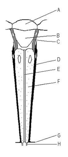
Os omatidios son tipicamente [[hexágono|hexafonais]] en sección transversal e aproximadamente dez veces máis longa que larga. O diámetro é maior na superficie, adelgazándose cara ao extremo interno. Na superficie exterior hai unha córnea, debaixo da cal hai un pseudocono que actúa enfocando máis a luz. A córnea e o pseudocono forman o 10% máis externo d lonxitude de todo o omatidio. |
|||
| ⚫ | |||
| ⚫ | O 90% máis interno do omatidio contén de 6 a 9 (dependendo da especie) [[célula fotorrecetora|células fotorreceptoras]] longas e delgadas no caso dalgunhas [[boolboreta]]s<ref name=briscoe2008>{{cite journal |pages=1805–13 |doi=10.1242/jeb.013045 |title=Reconstructing the ancestral butterfly eye: Focus on the opsins |year=2008 |last1=Briscoe |first1=A. D. |journal=Journal of Experimental Biology |volume=211 |issue=11 |pmid=18490396}}</ref> a miúdo abreviadas co nome de "[[célula R|células R]]" na literatura e xeralmente numeradas, por exemplo de R1 a R9.<ref name=briscoe2008/> Estas "células R" empaquetan estreitamente o omatidio. A porción de células R no eixe central do omatidio forman colectivamente unha guía para a luz, un tubo trnasparente, chamado '''rabdoma'''. |
||
| ⚫ | |||
In true [[fly|flies]], the rhabdom has separated into seven independent rhabdomeres (there are actually eight, but the two central rhabdomeres responsible for color vision sit one atop the other), such that a small inverted 7-pixel image is formed in each ommatidium. Simultaneously, the rhabdomeres in adjacent ommatidia are aligned such that the field of view within an ommatidium is the same as that between ommatidia. The advantage of this arrangement is that the same visual axis is sampled from a larger area of the eye, thereby increasing sensitivity by a factor of seven, without increasing the size of the eye or reducing its acuity. Achieving this has also required the rewiring of the eye such that the axon bundles are twisted through 180 degrees (re-inverted), and each rhabdomere is united with those from the six adjacent ommatidia that share the same visual axis. Thus, at the level of the lamina - the first optical processing center of the [[insect brain]] - the signals are input in exactly the same manner as in the case of a normal apposition compound eye, but the image is enhanced. This visual arrangement is known as ''neural superposition''.<ref>Land, Michael F. and Nilsson, Dan-Eric. ''Animal Eyes'', Second Edition. Oxford University Press, 2012. pp. 163-164. {{ISBN|978-0-19-958114-6}}.</ref> |
In true [[fly|flies]], the rhabdom has separated into seven independent rhabdomeres (there are actually eight, but the two central rhabdomeres responsible for color vision sit one atop the other), such that a small inverted 7-pixel image is formed in each ommatidium. Simultaneously, the rhabdomeres in adjacent ommatidia are aligned such that the field of view within an ommatidium is the same as that between ommatidia. The advantage of this arrangement is that the same visual axis is sampled from a larger area of the eye, thereby increasing sensitivity by a factor of seven, without increasing the size of the eye or reducing its acuity. Achieving this has also required the rewiring of the eye such that the axon bundles are twisted through 180 degrees (re-inverted), and each rhabdomere is united with those from the six adjacent ommatidia that share the same visual axis. Thus, at the level of the lamina - the first optical processing center of the [[insect brain]] - the signals are input in exactly the same manner as in the case of a normal apposition compound eye, but the image is enhanced. This visual arrangement is known as ''neural superposition''.<ref>Land, Michael F. and Nilsson, Dan-Eric. ''Animal Eyes'', Second Edition. Oxford University Press, 2012. pp. 163-164. {{ISBN|978-0-19-958114-6}}.</ref> |
||
Revisión como estaba o 23 de xuño de 2018 ás 21:45
Este artigo está a ser traducido ao galego por un usuario desta Wikipedia; por favor, non o edite. O usuario Miguelferig (conversa · contribucións) realizou a última edición na páxina hai 5 anos. Se o usuario non publica a tradución nun prazo de trinta días, procederase ó seu borrado rápido. |


Os 'omatidios son as unidades das que se compón o ollo composto dos artrópodos, como insectos, crustáceos e miriápodos[1] Un omatidio contén un grupo de células fotorreceptoras rodeadas por células de sostén e células pigmentadas. A parte externa do omatidio está cuberta por unha córnea transparente. Cada omatidio está inervado por un feixe de axóns (que xeralmente consta de 6 a 9 axóns, dependendo do número de rabdómeros)[2] e proporciona ao cerebro un elemento ou punto da imaxe (píxel). O cerebro forma unha imaxe conxunta a partir deses elementos de imaxe independentes. O número de omatidios no ollo depende do tipo de artrópodo e pode ser de só 5, como no isópodo antártico Glyptonotus antarcticus,[3] ou variar de só uns poucos no primitivo Zygentoma a arredor de 30 000 en grandes libélulas anisópteros como nalgunhas avelaíñas esfínxidas.[4]
Os omatidios son tipicamente hexafonais en sección transversal e aproximadamente dez veces máis longa que larga. O diámetro é maior na superficie, adelgazándose cara ao extremo interno. Na superficie exterior hai unha córnea, debaixo da cal hai un pseudocono que actúa enfocando máis a luz. A córnea e o pseudocono forman o 10% máis externo d lonxitude de todo o omatidio.
O 90% máis interno do omatidio contén de 6 a 9 (dependendo da especie) células fotorreceptoras longas e delgadas no caso dalgunhas boolboretas[5] a miúdo abreviadas co nome de "células R" na literatura e xeralmente numeradas, por exemplo de R1 a R9.[5] Estas "células R" empaquetan estreitamente o omatidio. A porción de células R no eixe central do omatidio forman colectivamente unha guía para a luz, un tubo trnasparente, chamado rabdoma.
Notas
- ↑ Muller, C; Sombke, A; Rosenberg, J (2007). "The fine structure of the eyes of some bristly millipedes (Penicillata, Diplopoda): Additional support for the homology of mandibulate ommatidia". Arthropod Structure & Development 36 (4): 463–76. doi:10.1016/j.asd.2007.09.002.
- ↑ Land, Michael F. and Nilsson, Dan-Eric. Animal Eyes, Second Edition. Oxford University Press, 2012. p. 162. ISBN 978-0-19-958114-6.
- ↑ Meyer-Rochow, Victor Benno. "The divided eye of the isopod Glyptonotus antarcticus: effects of unilateral dark adaptation and temperature elevation". Proceedings of the Royal Society of London B215: 433–450.
- ↑ Common, I. F. B. (1990). Moths of Australia. Brill. p. 15. ISBN 978-90-04-09227-3.
- ↑ 5,0 5,1 Briscoe, A. D. (2008). "Reconstructing the ancestral butterfly eye: Focus on the opsins". Journal of Experimental Biology 211 (11): 1805–13. PMID 18490396. doi:10.1242/jeb.013045.
