
Ficheiro:GFP Superresolution Christoph Cremer.JPG
GFP_Superresolution_Christoph_Cremer.JPG (538 × 389 píxeles; tamaño do ficheiro: 156 kB; tipo MIME: image/jpeg)
| Este ficheiro procede de Wikimedia Commons. A continuación móstrase a información da súa páxina de descrición. Commons é un repositorio libre de ficheiros multimedia. Pode contribuír alí cargando as súas imaxes. |
Resumo
| Descrición |
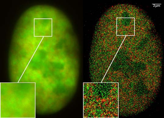
GFP superresolution, optical nanoscopy ( Christoph Cremer, emeritus at Heidelberg university [1]) View of a nucleus of a bone cancer cell: using normal high resolution fluorescence microscopy, it is not possible to distinguish details of its structure (image on the left). Using the two Color Localization Microscopy 2CLM (image on the right) it is possible to localize 70,000 histone molecules (red: RFP-H2A) and 50,000 chromatin remodeling proteins (green: GPF-Snf2H) in a field of view of 470 µm2 with an optical depth of 600 nm. Common fluorescence markers were used. 2CLM is the only optical nanoscopy method that allows position based co-localization of single molecules at high density in a wide field of view using conventional fluorescent proteins such as GFP, YFP, RFP, or other conventional fluorochromes. Due to its high optical single molecule resolution, 2CLM allows significantly more precise analyses of potential protein interactions than FRET-(Fluorescence Resonance Energy Transfer) technology, which is at present the preferred method for such investigations. This is of particular significance in studies of biomolecular machines (BMMs) within cells: Single BMMS can be analysed, including the number of molecules of a given type; distances between proteins in these BMMs often are substantially greater than those that can be analyzed by FRET (restricted to a maximum distance of only a few nm). Possible to use conventional, well established and inexpensive fluorescent dyes, from the GFP group, and its dye variants, to the well-known Alexa and fluorescein dyes. Fundamental to SPDMphymod are blinking phenomena (flashes of fluorescence), induced by reversible bleaches (metastable dark states). Individual molecules of the same spectral emission color can be detected. Publikation: Manuel Gunkel, Fabian Erdel, Karsten Rippe, Paul Lemmer, Rainer Kaufmann, Christoph Hörmann, Roman Amberger and Christoph Cremer: Dual color localization microscopy of cellular nanostructures. In: Biotechnology Journal, 2009, 4, 927-938. ISSN 1860-6768 |
| Data | 073009 |
| Orixe | Obra propia |
| Autoría | Andy Nestl |
| Licenza (Reuso deste ficheiro) |
Gallery
- Super Resolution Microscopy - Localisation Microscopy
-
 Breast Cancer Cells: 3D Dual Color Super Resolution Microscopy of Her2 and Her3 & cluster calculations
 -
 Single YFP molecule detection in a human cancer cell. Typical distance measurements 15 nm
 -
 Co- localisation microscopy with GFP and RFP fusion proteins (nucleus of a bone cancer cell) 120.000 localized molecules in a widefield area(470 µm2)
 -
 Label-free Localisation Microscopy SPDM - Super Resolution Microscopy reveals prior undetebable intracellular structures
 -
 Investigation of human eye tissue, affected by macular degeneration AMD
 -
 Virus Super Resolution Microscopy SPDM Cremer/Wege labs


Licenza
- Vostede é libre de:
- compartir – copiar, distribuír e difundir a obra
- facer obras derivadas – adaptar a obra
- Baixo as seguintes condicións:
- recoñecemento – Debe indicar a debida atribución de autoría, fornecer unha ligazón á licenza e indicar se se realizaron cambios. Pode facer isto de calquera forma razoable, mais non nunha forma que indique que quen posúe a licenza apoia ou subscribe o seu uso da obra.
- compartir igual – Se altera, transforma ou amplía este contido, debe publicar as súas contribucións baixo a mesma licenza ou outra compatible á orixinal.

|
Autorízase a copia, distribución e/ou modificación deste documento baixo os termos da licenza de documentación libre GNU, versión 1.2 ou calquera outra que posteriormente publique a Free Software Foundation; sen seccións invariables, textos de portada, nin textos de contraportada. Inclúese unha copia da devandita licenza na sección titulada GNU Free Documentation License. |
Resumo
- ↑ https://www.physik.uni-heidelberg.de/personen/lsf.php?details=1537 |titel=Fakultät für Physik und Astronomie |abruf=2020-10-01
Historial do ficheiro
Prema nunha data/hora para ver o ficheiro tal e como estaba nese momento.
| Data/Hora | Miniatura | Dimensións | Usuario | Comentario | |
|---|---|---|---|---|---|
| actual | 30 de xullo de 2009 ás 12:14 | | 538 × 389 (156 kB) | Andy Nestl | {{Information |Description=GFP superresolution, optical nanoscopy (Christoph Cremer) |Source=Own work by uploader |Date=073009 |Author=Andy Nestl |Permission=given by Christoph Cremer, University of Heidelberg |other_versions= }} |
Uso do ficheiro
As seguintes 2 páxinas usan este ficheiro:
Uso global do ficheiro
Os seguintes wikis empregan esta imaxe:
- Uso en ar.wikipedia.org
- Uso en be.wikipedia.org
- Uso en bn.wikipedia.org
- Uso en ca.wikipedia.org
- Uso en cs.wikipedia.org
- Uso en de.wikipedia.org
- Uso en en.wikipedia.org
- Uso en en.wikibooks.org
- Uso en eo.wikipedia.org
- Uso en fa.wikipedia.org
- Uso en fr.wikipedia.org
- Uso en he.wikipedia.org
- Uso en it.wikipedia.org
- Uso en mai.wikipedia.org
- Uso en ne.wikipedia.org
- Uso en nl.wikipedia.org
- Uso en pl.wikipedia.org
- Uso en pt.wikipedia.org
- Uso en sv.wikipedia.org
- Uso en ta.wikipedia.org
- Uso en vi.wikipedia.org
- Uso en zh.wikipedia.org