
Ficheiro:Phototransduction.png
Tamaño desta vista previa: 800 × 338 píxeles. Outras resolucións: 320 × 135 píxeles | 640 × 271 píxeles | 1.327 × 561 píxeles.
Ficheiro orixinal (1.327 × 561 píxeles; tamaño do ficheiro: 344 kB; tipo MIME: image/png)
| Este ficheiro procede de Wikimedia Commons. A continuación móstrase a información da súa páxina de descrición. Commons é un repositorio libre de ficheiros multimedia. Pode contribuír alí cargando as súas imaxes. |
| Descrición |
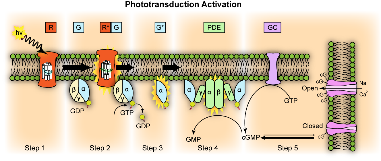
English: Representation of molecular steps in photoactivation (modified from Leskov et al., 2000). Depicted is an outer membrane disk in a rod. Step 1: Incident photon (hv) is absorbed and activates a rhodopsin by conformational change in the disk membrane to R*. Step 2: Next, R* makes repeated contacts with transducin molecules, catalyzing its activation to G* by the release of bound GDP in exchange for cytoplasmic GTP. The α and γ subunits Step 3: G* binds inhibitory γ subunits of the phosphodiesterase (PDE) activating its α and β subunits. Step 4: Activated PDE hydrolyzes cGMP. Step 5: Guanylyl cyclase (GC) synthesizes cGMP, the second messenger in the phototransduction cascade. Reduced levels of cytosolic cGMP cause cyclic nucleotide gated channels to close preventing further influx of Na+ and Ca2+.
Deutsch: Repräsentation der molekularen Schritte der Lichtaktivierung (verändert nach Leskov et al., 2000). Es wird die äussere Membranschiebe eines Stäbchens abgebildet.
|
||
| Data | |||
| Orixe | http://en.wikipedia.org/wiki/File:Phototransduction.png | ||
| Autoría | Jason J. Corneveaux, wiki user: Caddymob (talk) | ||
| Licenza (Reuso deste ficheiro) |
Eu, como posuidor dos dereitos de autor desta obra, pola presente publícoa baixo as seguintes licenzas:
Este ficheiro está licenciado baixo a licenza Creative Commons recoñecemento 3.0 Unported.
Pode seleccionar a licenza que desexe. |
||
| Outras versións | Obras derivadas deste ficheiro: Phototransduction uk.png |

Historial do ficheiro
Prema nunha data/hora para ver o ficheiro tal e como estaba nese momento.
| Data/Hora | Miniatura | Dimensións | Usuario | Comentario | |
|---|---|---|---|---|---|
| actual | 18 de abril de 2010 ás 15:42 | | 1.327 × 561 (344 kB) | Thomas.haslwanter | {{Information |Description=Representation of molecular steps in photoactivation (modified from Leskov et al., 2000). Depicted is an outer membrane disk in a rod. Step 1: Incident photon (hv) is absorbed and activates a rhodopsin by conformational change i |
Uso do ficheiro
A seguinte páxina usa este ficheiro:
Uso global do ficheiro
Os seguintes wikis empregan esta imaxe:
- Uso en bg.wikibooks.org
- Uso en de.wikibooks.org
- Uso en el.wikibooks.org
- Uso en en.wikipedia.org
- Uso en en.wikibooks.org
- Uso en et.wikipedia.org
- Uso en eu.wikipedia.org
- Uso en fa.wikipedia.org
- Uso en fr.wikibooks.org
- Uso en it.wikibooks.org
- Uso en ml.wikipedia.org
- Uso en mn.wikipedia.org
- Uso en pt.wikibooks.org