
Ficheiro:HIV-budding-Color.jpg

Tamaño desta vista previa: 800 × 531 píxeles. Outras resolucións: 320 × 213 píxeles | 640 × 425 píxeles | 1.024 × 680 píxeles | 1.280 × 850 píxeles | 2.967 × 1.971 píxeles.
Ficheiro orixinal (2.967 × 1.971 píxeles; tamaño do ficheiro: 3,92 MB; tipo MIME: image/jpeg)
| Este ficheiro procede de Wikimedia Commons. A continuación móstrase a información da súa páxina de descrición. Commons é un repositorio libre de ficheiros multimedia. Pode contribuír alí cargando as súas imaxes. |
Resumo
| Descrición |
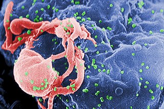
English: Scanning electron micrograph of HIV-1 budding (in green) from cultured lymphocyte. This image has been colored to highlight important features; see PHIL 1197 for original black and white view of this image.
Multiple round bumps on cell surface represent sites of assembly and budding of virions.
Español: Microfotografía con MEB de VIH-1 en liberación (en verde) en un cultivo de linfocitos. Esta imagen ha sido coloreada para resaltar las características importantes; para la imagen original en blanco y negro véase PHIL 1197. Las múltiples protuberancias redondeadas sobre la superficie celular representa los sitios de ensamblado y gemación de viriones.
Français : Virus HIV fixé sur un lymphocyte vu en microscopie électronique (fausses couleurs, le VIH est en vert).
Bahasa Indonesia: HIV yang baru memperbanyak diri tampak bermunculan sebagai bulatan-bulatan kecil (diwarnai hijau) pada permukaan limfosit setelah menyerang sel tersebut; dilihat dengan mikroskop elektron.
Русский: Фотография, полученная с помощью сканирующего электронного микроскопа. Вирусы ВИЧ (зелёные) отпочковываются от заражённого лимфоцита. Фотография была раскрашена с целью подчеркнуть важные детали; см. исходную чёрно-белую версию ниже.
Многочисленные круглые выпуклости на поверхности клетки являются местами сборки и отпочковывания вирионов.
Български: Вирусът ХИВ (в зелено) разспространяващ се от вече заразен лимфоцит.
Polski: Fotografia wykonana skaningowym mikroskopem elektronowym - przedstawia wirusy (kolor zielony) wydostających się z limfocytu. |
||
| Data | |||
| Orixe |
|
||
| Autoría |
|
||
| Licenza (Reuso deste ficheiro) |
PD-USGov-HHS-CDC English: None - This image is in the public domain and thus free of any copyright restrictions. As a matter of courtesy we request that the content provider be credited and notified in any public or private usage of this image. |
||
| Outras versións |
|


fuk12
Licenza
Esta imagem é um trabalho dos Centers for Disease Control and Prevention, parte do Departamento de Saúde e Serviços Humanos dos Estados Unidos da América, tirada ou feita durante o curso de uma tarefa oficial de um funcionário. Como trabalho do Governo Federal dos Estados Unidos da América, a imagem está no domínio público.
|
Historial do ficheiro
Prema nunha data/hora para ver o ficheiro tal e como estaba nese momento.
| Data/Hora | Miniatura | Dimensións | Usuario | Comentario | |
|---|---|---|---|---|---|
| actual | 20 de abril de 2008 ás 00:16 | | 2.967 × 1.971 (3,92 MB) | Optigan13 | {{Information |Description={{en|Scanning electron micrograph of HIV-1 budding from cultured lymphocyte. See PHIL 1197 for a black and white view of this image. Multiple round bumps on cell surface represent sites of assembly and budding of virions.}} |Sou |
Uso do ficheiro
As seguintes 3 páxinas usan este ficheiro:
Uso global do ficheiro
Os seguintes wikis empregan esta imaxe:
- Uso en ar.wikipedia.org
- Uso en arz.wikipedia.org
- Uso en ast.wikipedia.org
- Uso en as.wikipedia.org
- Uso en azb.wikipedia.org
- Uso en az.wikipedia.org
- Uso en be-tarask.wikipedia.org
- Uso en bg.wikipedia.org
- Uso en bn.wikipedia.org
- Uso en ca.wikipedia.org
- Uso en ca.wikinews.org
- Uso en ckb.wikipedia.org
- Uso en cs.wikipedia.org
- Wikipedie:Studenti píší Wikipedii/Pokroky v imunologii I (2013/2014)
- Wikipedie:Studenti píší Wikipedii/Pokroky v imunologii I (2014/2015)
- Wikipedie:Nástěnka/Univerzita Karlova/Pokroky v imunologii (2013-2014)
- Wikipedie:Nástěnka/Univerzita Karlova/Molekulární imunologie (2014-2015)
- Wikipedie:Nástěnka/Univerzita Karlova/Pokroky v imunologii (2014-2015)
- Uso en cy.wikipedia.org
- Uso en de.wikipedia.org
- Uso en diq.wikipedia.org
- Uso en en.wikipedia.org
- Uso en en.wikibooks.org
Ollar o uso global deste ficheiro.