
Ficheiro:Electron micrograph of neuromuscular junction (cross-section).jpg
Non se pode ver nunha resolución meirande.
Electron_micrograph_of_neuromuscular_junction_(cross-section).jpg (433 × 289 píxeles; tamaño do ficheiro: 95 kB; tipo MIME: image/jpeg)
| Este ficheiro procede de Wikimedia Commons. A continuación móstrase a información da súa páxina de descrición. Commons é un repositorio libre de ficheiros multimedia. Pode contribuír alí cargando as súas imaxes. |
.jpg)
Resumo
| Descrición |
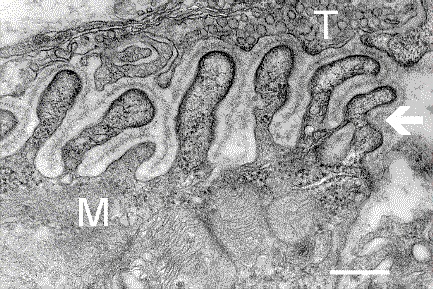
English: Electron micrograph showing a cross-section through the neuromuscular junction. T is the axon terminal, M is the muscle fiber. The arrow shows junctional folds with basal lamina. Postsynaptic densities are visible on the tips between the folds. The scale is 0.3 µm. |
| Data | Originally uploaded to en.wikipedia on 10 de marzo de 2006. |
| Orixe | Synapse Web at the National Institute of Mental Health, National Institutes of Health; originally from en.wikipedia; description page is/was here. |
| Autoría | National Institute of Mental Health; originally uploaded by Nrets at en.wikipedia. |
Licenza
This image is a work of the National Institutes of Health, part of the United States Department of Health and Human Services, taken or made as part of an employee's official duties. As a work of the U.S. federal government, the image is in the public domain.
 |
||
| Este ficheiro foi identificado como libre de restricións baixo as leis de dereitos de autor, incluídos todos os dereitos relacionados. | ||
Rexistro de cargas orixinal
(All user names refer to en.wikipedia)
- 2006-03-10 20:07 Nrets 433×289×8 (97758 bytes) Electron micrograph showing a cross section through the neuromuscular junction. T is the axon terminal, M is the muscle fiber. The arrow shows junctional folds with basal lamina. Postsynaptic densities are visible on the tips between the folds. Scale is 0
Historial do ficheiro
Prema nunha data/hora para ver o ficheiro tal e como estaba nese momento.
| Data/Hora | Miniatura | Dimensións | Usuario | Comentario | |
|---|---|---|---|---|---|
| actual | 22 de marzo de 2007 ás 03:41 | | 433 × 289 (95 kB) | Fran Rogers | {{Information |Description=Electron micrograph showing a cross section through the neuromuscular junction. T is the axon terminal, M is the muscle fiber. The arrow shows junctional folds with basal lamina. Postsynaptic densities are visible on the tips be |
Uso do ficheiro
A seguinte páxina usa este ficheiro:
Uso global do ficheiro
Os seguintes wikis empregan esta imaxe:
- Uso en ar.wikipedia.org
- Uso en cs.wikipedia.org
- Uso en de.wikipedia.org
- Uso en en.wikipedia.org
- Uso en es.wikipedia.org
- Uso en fa.wikipedia.org
- Uso en he.wikipedia.org
- Uso en ko.wikipedia.org
- Uso en pt.wikipedia.org
- Uso en ru.wikipedia.org
- Uso en uk.wikipedia.org
- Uso en zh.wikipedia.org
.jpg)