
Membrana basilar: Diferenzas entre revisións
Sen resumo de edición |
Sen resumo de edición |
||
| Liña 5: | Liña 5: | ||
| GraySubject = 232 |
| GraySubject = 232 |
||
| GrayPage = 1056 |
| GrayPage = 1056 |
||
| Imaxe = Organ_of_corti.svg |
| Imaxe = Organ_of_corti gl.svg |
||
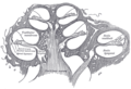
| Lenda = Sección a ravés do [[órgano de Corti]], na que se mostra a membrana basilar |
| Lenda = Sección a ravés do [[órgano de Corti]], na que se mostra a membrana basilar |
||
| Imaxe2 = Cochlea-crosssection gl.svg |
| Imaxe2 = Cochlea-crosssection gl.svg |
||
Revisión como estaba o 25 de agosto de 2015 ás 17:38
 | Este artigo está a ser traducido ao galego por un usuario desta Wikipedia; por favor, non o edite. O usuario Miguelferig (conversa · contribucións) realizou a última edición na páxina hai 8 anos. Se o usuario non publica a tradución nun prazo de trinta días, procederase ó seu borrado rápido. |
| Membrana basilar | |
|---|---|
 | |
| Sección a ravés do órgano de Corti, na que se mostra a membrana basilar | |
 | |
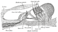
| Sección transversal da cóclea. | |
| Latín | membrana basilaris ductus cochlearis |
| Gray's | pág.1056 |
| MeSH | Basilar+membrane |
A membrana basilar situada na cóclea do oído interno é un elemento estrutural ríxido que separa os dous tubos ou escalas cheas de líquidos que corren en espiral ao longo da cóclea, que son a escala media e a escala timpática (ver figura).
Estrutura
The basilar membrane is a pseudo-resonant structure[1] that, like strings on an instrument, varies in width and stiffness. The "string" of the basilar membrane is not a set of parallel strings, as in a guitar, but a long structure that has different properties (width, stiffness, mass, damping, and the dimensions of the ducts that it couples to) at different points along its length. The motion of the basilar membrane is generally described as a traveling wave.[2] The parameters of the membrane at a given point along its length determine its characteristic frequency (CF), the frequency at which it is most sensitive to sound vibrations. The basilar membrane is widest (0.42–0.65 mm) and least stiff at the apex of the cochlea, and narrowest (0.08–0.16 mm) and most stiff at the base.[3] High-frequency sounds localize near the base of the cochlea (near the round and oval windows), while low-frequency sounds localize near the apex.
Función

Separación endolinfa/perilinfa
The fluids in these two tubes, the endolymph and the perilymph are very different chemically, biochemically, and electrically. Therefore they are kept strictly separated. This separation is the main function of Reissner's membrane (between scala vestibuli and scala media), and it is also the function of tissue held by the basilar membrane such as the inner and outer sulcus cells (shown in yellow) and the reticular lamina of the organ of Corti (shown in magenta). For the organ of Corti the basilar membrane is permeable to perilymph. Here the border between endolymph and perilymph occurs at the reticular lamina, the endolymph side of the organ of Corti.[6]
Base para as células sensoriais
The basilar membrane is also the base for the sensory cells of hearing, the hair cells that are equipped with "Stereocilia". There are approximately 15,000 hair cells in each human ear (see figure). This function as base of the sensory cells gave the basilar membrane its name, and it is again present in all land vertebrates. Due to its location, the basilar membrane places the hair cells in a position where they are adjacent to both the endolymph and the perilymph, which is a precondition of hair cell function.
Dispersión de frecuencias
A third, evolutionarily younger, function of the basilar membrane is strongly developed in the cochlea of most mammalian species and weakly developed in some bird species:[7] the dispersion of incoming sound waves to separate frequencies spatially. In brief, the membrane is tapered and it is stiffer at one end than at the other. Furthermore, sound waves travelling to the far, "floppier" end of the basilar membrane have to travel through a longer fluid column than sound waves travelling to the nearer, stiffer end. Each part of the basilar membrane, together with the surrounding fluid, can therefore be thought of as a "mass-spring" system with different resonant properties: high stiffness and low mass, hence high resonant frequencies at the near end, and low stiffness and high mass, hence low resonant frequencies, at the far end.[8] This causes sound input of a certain frequency to vibrate some locations of the membrane more than other locations. As shown in experiments by Nobel Prize laureate Georg von Békésy, high frequencies lead to maximum vibrations at the basal end of the cochlear coil, where the membrane is narrow and stiff, and low frequencies lead to maximum vibrations at the apical end of the cochlear coil, where the membrane is wider and more compliant. This "place–frequency map" can be described quantitatively by the Greenwood function and its variants.
Sound-driven vibrations travel as waves along this membrane, along which, in humans, lie about 3,500 inner hair cells spaced in a single row. Each cell is attached to a tiny triangular frame. The 'hairs' are minute processes on the end of the cell, which are very sensitive to movement. When the vibration of the membrane rocks the triangular frames, the hairs on the cells are repeatedly displaced, and that produces streams of corresponding pulses in the nerve fibers, which are transmitted to the auditory pathway.[9] The outer hair cells feed back energy to amplify the traveling wave, by up to 65 dB at some locations.[10][11]
Galería
-
 Sección lon xitudinal da cóclea.
 -
 Piso do conduto coclear.
 -
 Limbo espiral e membrana basilar.
 -
 Sección a través do órgano espiral de Corti (ampliada).
 -
 A membrana reticular e estruturas subxacentes.


Notas
- ↑ M. Holmes and J. D. Cole, "Pseudoresonance in the cochlea, ' in: Mechanics of Hearing, E. de Boer and M. A. Viergever (editors), Proceedings of the IUTAM/ICA Symposium, Delft (1983), pp. 45-52.
- ↑ Richard R. Fay, Arthur N. Popper, and Sid P. Bacon (2004). Compression: From Cochlea to Cochlear Implants. Springer. ISBN 0-387-00496-3.
- ↑ Oghalai JS. The cochlear amplifier: augmentation of the traveling wave within the inner ear. Current Opinion in Otolaryngology & Head & Neck Surgery. 12(5):431-8, 2004
- ↑ Shera, Christopher A. (2007). "Laser amplification with a twist: Traveling-wave propagation and gain functions from throughout the cochlea". Journal of the Acoustical Society of America 122 (5): 2738–2758. doi:10.1121/1.2783205. Consultado o 13 April 2013.
- ↑ Robles, L.; Ruggero, M. A. (2001). "Mechanics of the mammalian cochlea". Physiological Reviews 81 (3): 1305–1352. Consultado o 13 April 2013.
- ↑ Salt, A.N., Konishi, T., 1986. The cochlear fluids: Perilymph and endolymph. In: Altschuler, R.A., Hoffman, D.W., Bobbin, R.P. (Eds.), Neurobiology of Hearing: The Cochlea. Raven Press, New York, pp. 109-122
- ↑ Fritzsch B: The water-to-land transition: Evolution of the tetrapod basilar papilla; middle ear, and auditory nuclei. In: Douglas B. Webster, Richard R. Fay, Arthur N. Popper, editors (1992). The Evolutionary biology of hearing. Berlin: Springer-Verlag. pp. 351–375. ISBN 0-387-97588-8.
- ↑ Schnupp J., Nelken I., King A. (2011). Auditory Neuroscience. Cambridge MA: MIT Press. ISBN 0-262-11318-X.
- ↑ Modelo:Cite document
- ↑ Nilsen KE, Russell IJ (1999). "Timing of cochlear feedback: spatial and temporal representation of a tone across the basilar membrane". Nat. Neurosci. 2 (7): 642–8. PMID 10404197. doi:10.1038/10197.
- ↑ Nilsen KE, Russell IJ (2000). "The spatial and temporal representation of a tone on the guinea pig basilar membrane". Proc. Natl. Acad. Sci. U.S.A. 97 (22): 11751–8. PMC 34345. PMID 11050205. doi:10.1073/pnas.97.22.11751.
Véxase tamén
Outros artigos
Ligazóns externas
- Auditory Neuroscience | The Ear con varias animacións que mostran o movemento da membrana basilar en varias condicións de estímulos
- Functional anatomy of the inner ear: con moitas imaxes, animacións, e explicacións moi concisas e funcionais
- Basilar Membrane Simulator Vídeo e textos sobre a estimulación da membrana basilar
- The role of the basilar membrane in sound reception: boa explicación e diagramas