
Ficheiro:MultiPhotonExcitation-Fig10-doi10.1186slash1475-925X-5-36.JPEG

Tamaño desta vista previa: 600 × 600 píxeles. Outras resolucións: 240 × 240 píxeles | 480 × 480 píxeles | 768 × 768 píxeles | 1.024 × 1.024 píxeles | 2.133 × 2.133 píxeles.
Ficheiro orixinal (2.133 × 2.133 píxeles; tamaño do ficheiro: 949 kB; tipo MIME: image/jpeg)
| Este ficheiro procede de Wikimedia Commons. A continuación móstrase a información da súa páxina de descrición. Commons é un repositorio libre de ficheiros multimedia. Pode contribuír alí cargando as súas imaxes. |
Resumo
| Descrición |
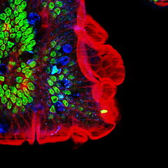
English: Original figure legend: Multiple fluorescence 2PE imaging. 2PE multiple fluorescence image from a 16 μm cryostat section of mouse intestine stained with a combination of fluorescent stains (F-24631, Molecular Probes). Alexa Fluor 350 wheat germ agglutinin, a blue-fluorescent lectin, was used to stain the mucus of goblet cells. The filamentous actin prevalent in the brush border was stained with red-fluorescent Alexa Flu or 568 phalloidin. Finally, the nuclei were stained with SYTOX ® Green nucleic acid stain. Imaging has been performed at 780 nm, 100 x 1.4 NA Leica objective, using a Chameleon XR ultrafast Ti-Sapphire laser (Coherent Inc., USA) coupled at LAMBS-MicroScoBio with a Spectral Confocal Laser Scanning Microscope, Leica SP2-AOBS.
Deutsch: Zweiphotonenaufnahme an einem Schnitt durch einen Mausdarm. Zellkerne in grün, Schleim der Becherzellen in blau, Aktin (Phalloidin-Färbung) in rot. Anregung erfolgte bei 780 nm durch einen Titan:Saphir-Laser. |
| Data | |
| Orixe |
Multi-photon excitation microscopy. BioMedical Engineering OnLine, 2006, 5:36. |
| Autoría | Alberto Diaspro, Paolo Bianchini, Giuseppe Vicidomini, Mario Faretta, Paola Ramoino and Cesare Usai |
| Licenza (Reuso deste ficheiro) |
Este ficheiro está licenciado baixo a licenza Creative Commons recoñecemento xenérico 2.0.
|
| Outras versións |
 |
All images uploaded from this article about multi-photon and two-photon-microscopy:


Historial do ficheiro
Prema nunha data/hora para ver o ficheiro tal e como estaba nese momento.
| Data/Hora | Miniatura | Dimensións | Usuario | Comentario | |
|---|---|---|---|---|---|
| actual | 23 de decembro de 2008 ás 18:59 | | 2.133 × 2.133 (949 kB) | Dietzel65 | == Beschreibung == {{Information |Description={{en|1=Original figure legend: ''Multiple fluorescence 2PE imaging. 2PE multiple fluorescence image from a 16 μm cryostat section of mouse intestine stained with a combination of fluorescent stains (F-24631, |
Uso do ficheiro
A seguinte páxina usa este ficheiro:
Uso global do ficheiro
Os seguintes wikis empregan esta imaxe:
- Uso en ar.wikipedia.org
- Uso en de.wikipedia.org
- Uso en en.wikipedia.org
- Uso en es.wikipedia.org
- Uso en fa.wikipedia.org
- Uso en fr.wikipedia.org
- Uso en ko.wikipedia.org
- Uso en pt.wikipedia.org
- Uso en sr.wikipedia.org